Research
Dissecting the biology of metastasis
More than 90% of cancer-related deaths are due to the development of a metastatic disease (external pageWHO). Worldwide, metastasis accounts for more than 7 million deaths each year, and little is known about how to suppress a metastatic disease in patients.
Our laboratory is interested in understanding the fundamental molecular mechanisms that drive cancer and its metastatic progression, with a particular focus on the analysis of circulating tumor cells. In our studies, we use a combination of molecular biology, next-generation sequencing, computational biology, microfluidic and robotic technologies, patient samples, in vivo models, genetic engineering, CRISPR screens and drug screens to better understand the biology and vulnerabilities of aggressive cancers. We collaborate actively with a number of academic research groups, hospitals, and healthcare companies throughout the world, striving to identify novel metastasis-relevant therapeutic opportunities to fight against the metastatic spread of cancer.
Circulating tumor cells and their clusters
Cancer cells that leave the primary tumor site and are transported through the circulation to distant organs are referred to as circulating tumor cells (CTCs). While CTCs are extraordinarily rare in the peripheral circulation of patients with cancer (approximately one CTC per billion normal blood cells), they hold the key to dissecting fundamental aspects of how metastasis occurs. We isolate CTC from the blood of cancer patients with the use of specialized microfluidic technologies, and interrogate them at the molecular and functional level to uncover their fundamental biological properties and vulnerabilities.
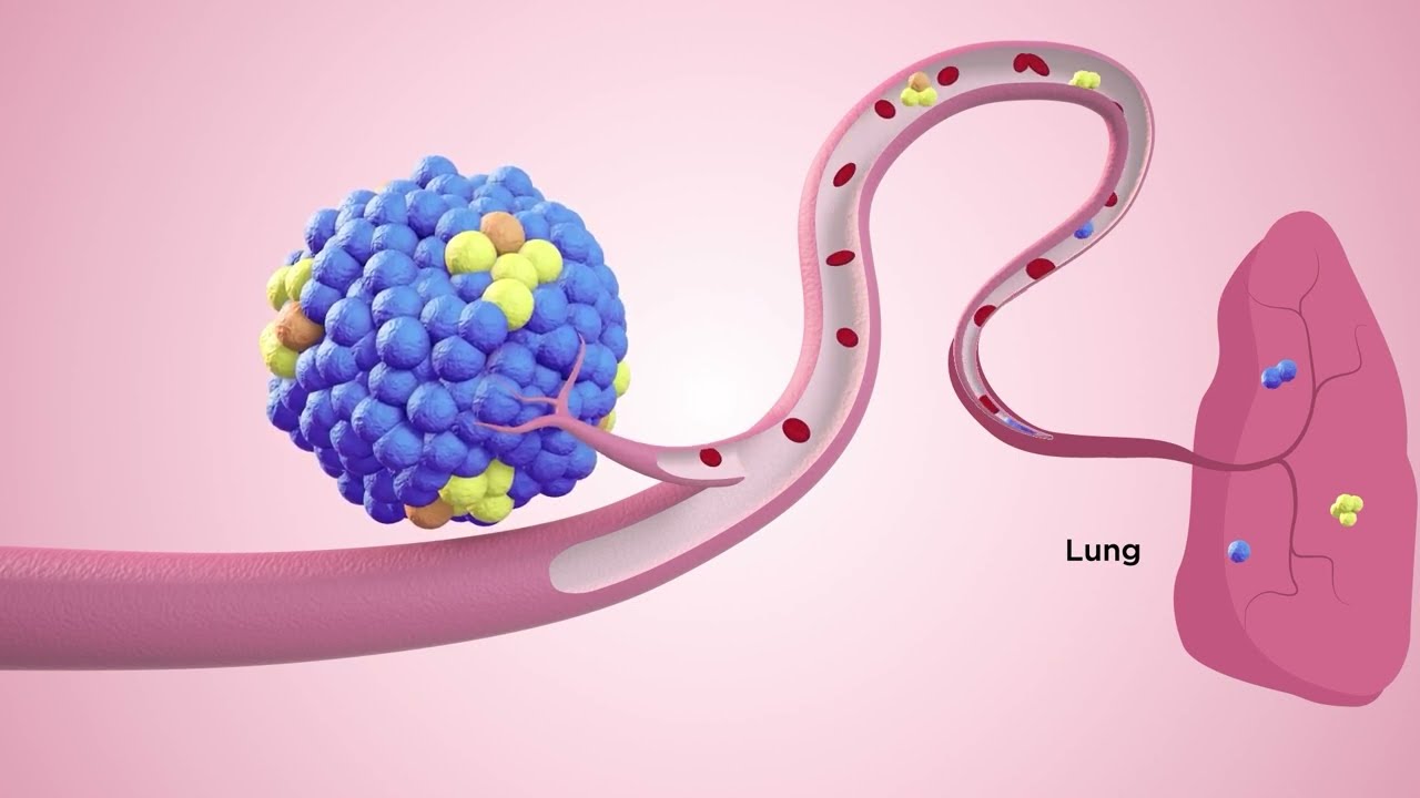
For example, we discovered a major role for CTC clusters (aggregates of cancer cells in circulation) in the metastatic process in breast cancer. We also found that CTC clusters are derivatives of hypoxic tumor areas and are characterized by hypomethylation of the binding sites for transcription factors that simultaneously regulate stemness and proliferation, such as OCT4, NANOG, SOX2 and SIN3A. Our results clearly link clustering and stemness features, and suggest that CTC cluster dissociation might be a valuable therapeutic strategy to reduce metastasis formation (external pageAceto et al., Cell, 2014; external pageGkountela et al, Cell, 2019; external pageDonato et al., Cell Rep, 2020) (Fig. 1).
We also discovered a new role for neutrophils in boosting the metastatic potential of CTCs. Particularly, we find that the association CTC-neutrophils enhances CTC proliferation through the release of specific cytokines. When this crosstalk is blocked, we observe a significant reduction in the metastatic spread of breast cancer cells (external pageSzczerba et al., Nature, 2019).
Together, our recent investigations on CTCs allowed us to better understand some of the key properties of the metastatic process, including the formation of homotypic and heterotypic CTC clusters, as well as some of their driving principles.
Timing and cues to circulating tumor cells intravasation
We are interested in understanding signals that dictate the intravasation of cancer cells in the bloodstream, as well as properties that define their ability to metastasize to various organs. A better understanding of these features may enable future therapeutic opportunities.
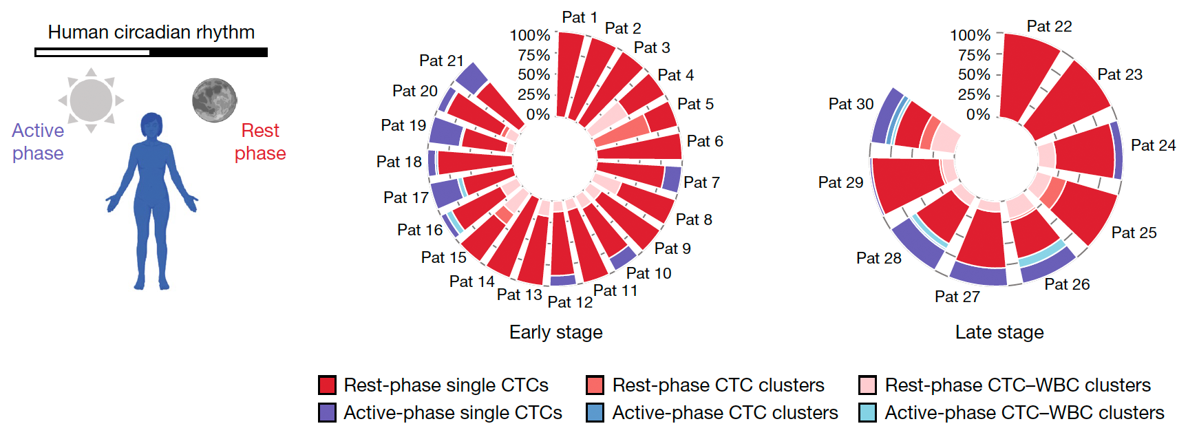
We discovered a key role of the circadian rhythm in regulating timing of CTC intravasation. Particularly, we observed that most CTC are generated during the rest phase (i.e. sleep) of breast cancer patients and mouse models, and that the majority of successful metastatic events can be ascribed to the same period. This occurs through the action of circadian rhythm-regulated hormones, which simultaneously regulate cancer cells proliferation and invasion into the bloodstream, yet in a time-dependent fashion (external pageDiamantopoulou et al., Nature, 2022) (Fig. 2).
Further, with a genome-wide CRISPR screen in vivo using patient-derived CTC xenografts, we improved our understanding of genes that regulate specific aspects of the metastatic cascade. These include genes that promote CTC intravasation and colonization to specific distant sites, such as bone, brain and liver (external pageScheidmann et al., Cancer Res, 2022).
Altogether, these findings allow us to plan future metastasis investigations in a time-controlled fashion, considering the role of the circadian rhythm in this process and well as the action of specific genes that are involved in this process in vivo.
Videos summary of our main discoveries, in a nutshell
Research in our lab is focused on improving our understanding of the metastatic process. Our key discoveries are summarized below, stay tuned for more…
2022
2014-2021
